Copyright
©The Author(s) 2017.
World J Orthop. Sep 18, 2017; 8(9): 697-704
Published online Sep 18, 2017. doi: 10.5312/wjo.v8.i9.697
Published online Sep 18, 2017. doi: 10.5312/wjo.v8.i9.697
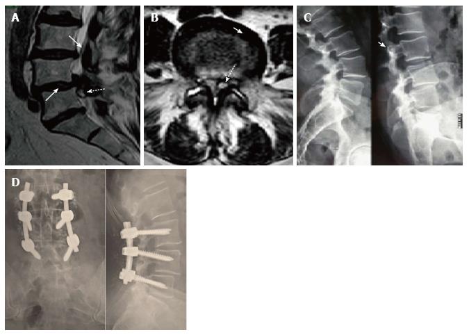
Figure 2 Case 12, Table 1.
Preoperative sagittal T2-weighted MR image (A) showing a spinal ganglion cyst (dotted arrow) accompanied by olisthesis at L4/L5 with a dehydrated intervertebral disk (arrow), partially herniated into the spinal canal. On axial images (B) the cyst (dotted arrow) appeared to be of the medium or articular type (see text for classification). The interfacetal space contained an anomalous abundance of “sinovia” (commonly called synovial fluid), as the contralateral one did. Dynamic X-rays (C) showed an unstable olisthesis at L4/L5 and L3/L4. Postoperative outcome of the L3/L5 stabilization is documented by standard X-ray films (D) which confirmed good stability and fusion of the lumbar spine.
- Citation: Domenicucci M, Ramieri A, Marruzzo D, Missori P, Miscusi M, Tarantino R, Delfini R. Lumbar ganglion cyst: Nosology, surgical management and proposal of a new classification based on 34 personal cases and literature review. World J Orthop 2017; 8(9): 697-704
- URL: https://www.wjgnet.com/2218-5836/full/v8/i9/697.htm
- DOI: https://dx.doi.org/10.5312/wjo.v8.i9.697


