Copyright
©The Author(s) 2017.
World J Orthop. Sep 18, 2017; 8(9): 697-704
Published online Sep 18, 2017. doi: 10.5312/wjo.v8.i9.697
Published online Sep 18, 2017. doi: 10.5312/wjo.v8.i9.697
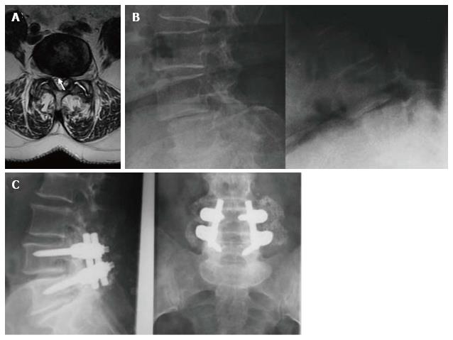
Figure 1 Case 2, Table 1.
Preoperative axial T2-weighted MR image (A) showing a dehydrated and hypointense disk with a hyperintense cystic formation at right L4-L5 level (arrow). The cyst appeared to be of the internal or flavum type (see text for the classification). Sagittal dynamic images (B) 12 mo after the first surgical treatment showed an unstable olisthesis at L4-L5 level. Standard X-rays performed 1 year after surgical stabilization (C) showed the instrumentation to be well-positioned with an optimal profile and fusion at L4-L5.
- Citation: Domenicucci M, Ramieri A, Marruzzo D, Missori P, Miscusi M, Tarantino R, Delfini R. Lumbar ganglion cyst: Nosology, surgical management and proposal of a new classification based on 34 personal cases and literature review. World J Orthop 2017; 8(9): 697-704
- URL: https://www.wjgnet.com/2218-5836/full/v8/i9/697.htm
- DOI: https://dx.doi.org/10.5312/wjo.v8.i9.697


