Copyright
©The Author(s) 2017.
World J Orthop. Sep 18, 2017; 8(9): 681-687
Published online Sep 18, 2017. doi: 10.5312/wjo.v8.i9.681
Published online Sep 18, 2017. doi: 10.5312/wjo.v8.i9.681
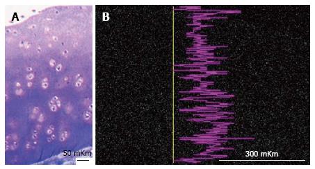
Figure 3 Articular cartilage of intact dog.
A: Semithin section, methylene blue stain. Ob. -6.3 х; oc. -12.5 х; B: Smart map shows sulphur distribution on scanned line. Instrumental magnification 170.
- Citation: Stupina T, Shchudlo M, Stepanov M. Electron probe microanalysis оf experimentally stimulated osteoarthrosis in dogs. World J Orthop 2017; 8(9): 681-687
- URL: https://www.wjgnet.com/2218-5836/full/v8/i9/681.htm
- DOI: https://dx.doi.org/10.5312/wjo.v8.i9.681


