Copyright
©The Author(s) 2017.
World J Orthop. Sep 18, 2017; 8(9): 660-673
Published online Sep 18, 2017. doi: 10.5312/wjo.v8.i9.660
Published online Sep 18, 2017. doi: 10.5312/wjo.v8.i9.660
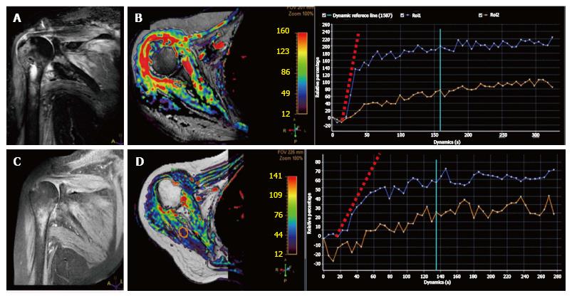
Figure 11 Magnetic resonance follow-up of septic arthritis in a 58-year-old woman with shoulder pain, fever, and swelling.
A: Coronal STIR shows severe edema within the proximal humerus as well as in the surrounding soft tissues with minimal intraarticular fluid; B: DCE-MRI relative enhancement map demonstrates extensive enhancement in the synovium and the humeral head; C, D: Follow-up MRI study 3 wk after intravenous antibiotic therapy demonstrates adequate response to treatment with reduction of edema on STIR and enhancement on DCE-MRI. Note also the change in the initial slope of the blue TIC (synovial enhancement) between pre and post-treatment studies. The orange TIC shows healthy muscle contrast enhancement as an internal reference. STIR: Short inversion time recovery; DWI: Diffusion-weighted imaging; MRI: Magnetic resonance imaging; DCE: Dynamic contrast enhancement.
- Citation: Martín Noguerol T, Luna A, Gómez Cabrera M, Riofrio AD. Clinical applications of advanced magnetic resonance imaging techniques for arthritis evaluation. World J Orthop 2017; 8(9): 660-673
- URL: https://www.wjgnet.com/2218-5836/full/v8/i9/660.htm
- DOI: https://dx.doi.org/10.5312/wjo.v8.i9.660


