Copyright
©The Author(s) 2017.
World J Orthop. Sep 18, 2017; 8(9): 660-673
Published online Sep 18, 2017. doi: 10.5312/wjo.v8.i9.660
Published online Sep 18, 2017. doi: 10.5312/wjo.v8.i9.660
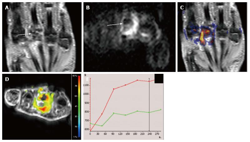
Figure 9 Multiparametric evaluation of rheumatoid arthritis in a 40-year-old woman with hand involvement.
A: Coronal STIR shows severe articular surface erosions with subchondral edema and synovial hypertrophy at the 3rd metatarsal-phalangeal joint (arrow); B: Axial DWI b800 demonstrates markedly restricted diffusion within this joint (arrow, B) with good correlation on (C) STIR and DWI b 800 fused image; D, E: (D) DCE-MRI relative enhancement map shows increased enhancement and (E) TIC of the involved joint (red curve) shows an initial fast enhancement which becomes more progressive and slow in late phases in comparison to the adjacent normal joint (green curve). STIR: Short inversion time recovery; DWI: Diffusion-weighted imaging; MRI: Magnetic resonance imaging; DCE: Dynamic contrast enhancement.
- Citation: Martín Noguerol T, Luna A, Gómez Cabrera M, Riofrio AD. Clinical applications of advanced magnetic resonance imaging techniques for arthritis evaluation. World J Orthop 2017; 8(9): 660-673
- URL: https://www.wjgnet.com/2218-5836/full/v8/i9/660.htm
- DOI: https://dx.doi.org/10.5312/wjo.v8.i9.660


