Copyright
©The Author(s) 2017.
World J Orthop. Sep 18, 2017; 8(9): 660-673
Published online Sep 18, 2017. doi: 10.5312/wjo.v8.i9.660
Published online Sep 18, 2017. doi: 10.5312/wjo.v8.i9.660
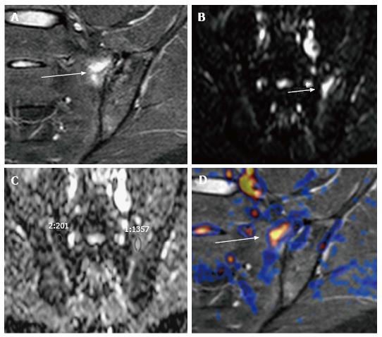
Figure 8 Acute sacroiliitis in a 32-year-old woman with left hip pain.
A-C: Coronal STIR shows a focus of subchondral bone edema in the left hemisacrum (arrow, A), which appears hyperintense on (B) high b value DWI, with (C) significantly higher ADC values than contralateral bone marrow (1.3 × 10⁻³ mm2/s vs 0.2 × 10⁻³ mm2/s); D: Fused DWI and STIR images allow a better depiction of bone edema. STIR: Short inversion time recovery; DWI: Diffusion-weighted imaging; ADC: Apparent diffusion coefficient.
- Citation: Martín Noguerol T, Luna A, Gómez Cabrera M, Riofrio AD. Clinical applications of advanced magnetic resonance imaging techniques for arthritis evaluation. World J Orthop 2017; 8(9): 660-673
- URL: https://www.wjgnet.com/2218-5836/full/v8/i9/660.htm
- DOI: https://dx.doi.org/10.5312/wjo.v8.i9.660


