Copyright
©The Author(s) 2017.
World J Orthop. Sep 18, 2017; 8(9): 660-673
Published online Sep 18, 2017. doi: 10.5312/wjo.v8.i9.660
Published online Sep 18, 2017. doi: 10.5312/wjo.v8.i9.660
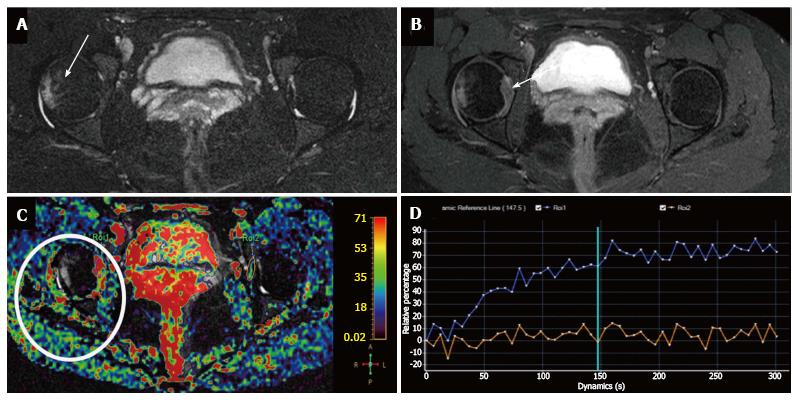
Figure 7 Evaluation of inflammatory arthritis with dynamic-contrast enhanced-magnetic resonance imaging in a 42-year-old woman with rheumatoid arthritis and right hip pain.
A: Axial STIR shows mild articular effusion in the right hip with subtle signs of subchondral bone edema (arrow); B: Axial post-gadolinium SPIR GE T1-weighted image demonstrates moderate synovial thickening and enhancement especially at the medial articular surface (arrow); C: Relative enhancement map; D: Dynamic-contrast enhanced-magnetic resonance imaging demonstrate a type I TIC (blue curve) in the right hip, with progressive enhancement, compared to absence of significant enhancement in the contralateral hip (orange curve). This finding helps to confirm the inflammatory involvement of right hip. STIR: Short inversion time recovery.
- Citation: Martín Noguerol T, Luna A, Gómez Cabrera M, Riofrio AD. Clinical applications of advanced magnetic resonance imaging techniques for arthritis evaluation. World J Orthop 2017; 8(9): 660-673
- URL: https://www.wjgnet.com/2218-5836/full/v8/i9/660.htm
- DOI: https://dx.doi.org/10.5312/wjo.v8.i9.660


