Copyright
©The Author(s) 2017.
World J Orthop. Sep 18, 2017; 8(9): 660-673
Published online Sep 18, 2017. doi: 10.5312/wjo.v8.i9.660
Published online Sep 18, 2017. doi: 10.5312/wjo.v8.i9.660
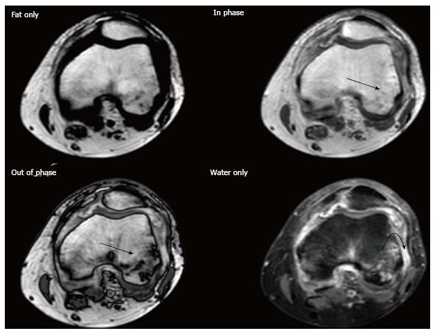
Figure 1 DIXON.
Thirty-two years old woman with knee pain and suspected rheumatoid arthritis. Post-contrast DIXON study was performed. Opposed-phase image shows large hypointense areas in both condyles (arrows), which are hardly seen on the in-phase image, consistent with bone marrow edema. Note the presence of bone erosions and synovitis (curved arrow) better depicted on water only imaging.
- Citation: Martín Noguerol T, Luna A, Gómez Cabrera M, Riofrio AD. Clinical applications of advanced magnetic resonance imaging techniques for arthritis evaluation. World J Orthop 2017; 8(9): 660-673
- URL: https://www.wjgnet.com/2218-5836/full/v8/i9/660.htm
- DOI: https://dx.doi.org/10.5312/wjo.v8.i9.660


