Copyright
©The Author(s) 2017.
World J Orthop. Aug 18, 2017; 8(8): 656-659
Published online Aug 18, 2017. doi: 10.5312/wjo.v8.i8.656
Published online Aug 18, 2017. doi: 10.5312/wjo.v8.i8.656
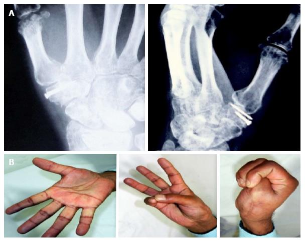
Figure 4 Clinico-radiological follow-up at 6 mo.
A: Radiograph at 6 mo follow up showing a healed fracture with maintainance of joint reduction; B: Clinical photograph showing good range of motion.
- Citation: Goyal T. Bennett’s fracture associated with fracture of Trapezium - A rare injury of first carpo-metacarpal joint. World J Orthop 2017; 8(8): 656-659
- URL: https://www.wjgnet.com/2218-5836/full/v8/i8/656.htm
- DOI: https://dx.doi.org/10.5312/wjo.v8.i8.656


