Copyright
©The Author(s) 2017.
World J Orthop. Aug 18, 2017; 8(8): 651-655
Published online Aug 18, 2017. doi: 10.5312/wjo.v8.i8.651
Published online Aug 18, 2017. doi: 10.5312/wjo.v8.i8.651
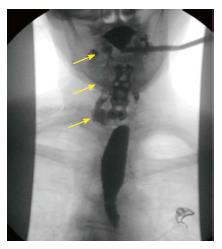
Figure 2 Anteroposterior fluoroscopic image of esophogram showed extravasated contrast material tracking along the right side of the neck (arrows).
- Citation: Elgafy H, Khan M, Azurdia J, Peters N. Open wound management of esophagocutaneous fistula in unstable cervical spine after corpectomy and multilevel laminectomy: A case report and review of the literature. World J Orthop 2017; 8(8): 651-655
- URL: https://www.wjgnet.com/2218-5836/full/v8/i8/651.htm
- DOI: https://dx.doi.org/10.5312/wjo.v8.i8.651


