Copyright
©The Author(s) 2017.
World J Orthop. Aug 18, 2017; 8(8): 606-611
Published online Aug 18, 2017. doi: 10.5312/wjo.v8.i8.606
Published online Aug 18, 2017. doi: 10.5312/wjo.v8.i8.606
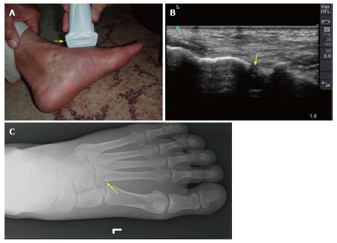
Figure 4 Point-of-care ultrasound of a 60-year-old man who twisted his left ankle and could not walk on it.
He had swelling of the left foot with maximum tenderness on the base of second metatarsal bone (A). The yellow arrow indicates the marker of linear probe which is shown on the left side of the screen while the groove on the other side is shown on the right side of the screen. B mode images of the previous patient showed a cortical defect (yellow arrow) at the base of the second metatarsal bone suggestive of a fracture (B). Plain X-ray of the foot confirmed these findings (C). Ultrasound study was performed by Professor Fikri Abu-Zidan, Department of Surgery, Al-Ain Hospital, Al-Ain, UAE.
- Citation: Abu-Zidan FM. Ultrasound diagnosis of fractures in mass casualty incidents. World J Orthop 2017; 8(8): 606-611
- URL: https://www.wjgnet.com/2218-5836/full/v8/i8/606.htm
- DOI: https://dx.doi.org/10.5312/wjo.v8.i8.606


