Copyright
©The Author(s) 2017.
World J Orthop. Aug 18, 2017; 8(8): 606-611
Published online Aug 18, 2017. doi: 10.5312/wjo.v8.i8.606
Published online Aug 18, 2017. doi: 10.5312/wjo.v8.i8.606
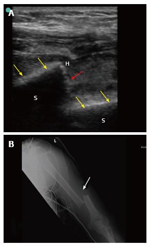
Figure 2 Point-of-care ultrasound of a 42-year-old laborer who fell from 8 meters high during work.
The patient presented with pain, swelling and deformity of the left arm. He had left wrist drop. B mode point-of-care ultrasound of the humeral shaft using a linear probe having a frequency of 10-12 MHz (A) showed that the white cortical line of the humeral shaft (yellow arrows) has been interrupted by a large gap (red arrow) suggesting a displaced fracture at the shaft. X-ray of the humerus (B) confirmed the presence of a displaced fracture (white arrow). S: Sonographic shadow of the humeral shaft; H: Hematoma at the edge of the fracture. Ultrasound study was performed by Professor Fikri Abu-Zidan, Department of Surgery, Al-Ain Hospital, Al-Ain, UAE.
- Citation: Abu-Zidan FM. Ultrasound diagnosis of fractures in mass casualty incidents. World J Orthop 2017; 8(8): 606-611
- URL: https://www.wjgnet.com/2218-5836/full/v8/i8/606.htm
- DOI: https://dx.doi.org/10.5312/wjo.v8.i8.606


