Copyright
©The Author(s) 2017.
World J Orthop. Aug 18, 2017; 8(8): 606-611
Published online Aug 18, 2017. doi: 10.5312/wjo.v8.i8.606
Published online Aug 18, 2017. doi: 10.5312/wjo.v8.i8.606
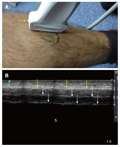
Figure 1 Ultrasound examination of the tibial shaft.
A linear probe having a frequency of 10-12 MHz was used (A). The marker (yellow arrow) is pointing proximally. The plain surface of the tibia makes the examination easy. Normal ultrasound findings (B) include a hyperechoic line (yellow arrows) representing the cortical line of the bone. There are reverberation artifacts deeper to this line (white arrows). These are linear lines parallel to the cortex, having the same distance between them and decreasing in density. A black shadow is located deeper to that. S: Sonographic shadow of the shaft of the tibia. Ultrasound study was performed by Professor Fikri Abu-Zidan, Department of Surgery, Al-Ain Hospital, Al-Ain, UAE.
- Citation: Abu-Zidan FM. Ultrasound diagnosis of fractures in mass casualty incidents. World J Orthop 2017; 8(8): 606-611
- URL: https://www.wjgnet.com/2218-5836/full/v8/i8/606.htm
- DOI: https://dx.doi.org/10.5312/wjo.v8.i8.606


