Copyright
©The Author(s) 2017.
World J Orthop. Jul 18, 2017; 8(7): 561-566
Published online Jul 18, 2017. doi: 10.5312/wjo.v8.i7.561
Published online Jul 18, 2017. doi: 10.5312/wjo.v8.i7.561
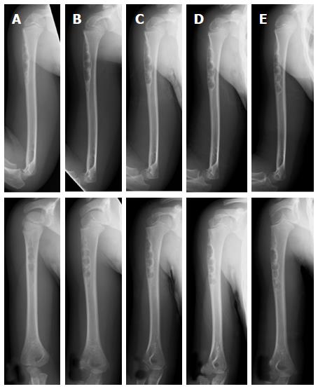
Figure 1 Plain radiographs of a non-ossifying fibroma in the humerus of a 4-year-old female.
An osteolytic lesion is shown at the cortex in the proximal humerus (A). Radiographs taken after 7 mo (B), 1 year (C), 1 year and 7 mo (D), and 2 years and 7 mo (E) together reveal the location of the lesion became more distal with growth of the child. The size of the lesion increased as it slowly ossified (plain radiographs: Anteroposterior view, top; lateral view, bottom).
- Citation: Sakamoto A, Arai R, Okamoto T, Matsuda S. Non-ossifying fibromas: Case series, including in uncommon upper extremity sites. World J Orthop 2017; 8(7): 561-566
- URL: https://www.wjgnet.com/2218-5836/full/v8/i7/561.htm
- DOI: https://dx.doi.org/10.5312/wjo.v8.i7.561


