Copyright
©The Author(s) 2017.
World J Orthop. Jun 18, 2017; 8(6): 484-490
Published online Jun 18, 2017. doi: 10.5312/wjo.v8.i6.484
Published online Jun 18, 2017. doi: 10.5312/wjo.v8.i6.484
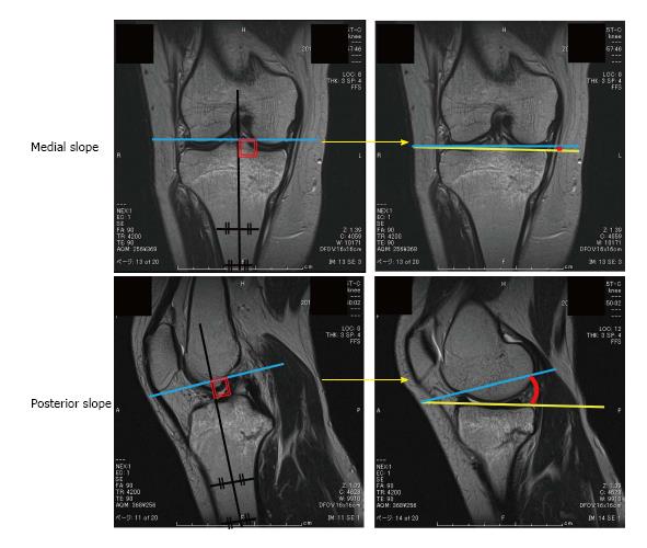
Figure 2 Magnetic resonance imaging illustrating the method used to determine the medial and posterior slopes.
The angles of tibial medial and posterior slopes were represented by a segment of red circle between blue and yellow lines.
- Citation: Yukata K, Yamanaka I, Ueda Y, Nakai S, Ogasa H, Oishi Y, Hamawaki JI. Medial tibial plateau morphology and stress fracture location: A magnetic resonance imaging study. World J Orthop 2017; 8(6): 484-490
- URL: https://www.wjgnet.com/2218-5836/full/v8/i6/484.htm
- DOI: https://dx.doi.org/10.5312/wjo.v8.i6.484


