Copyright
©The Author(s) 2017.
World J Orthop. May 18, 2017; 8(5): 379-384
Published online May 18, 2017. doi: 10.5312/wjo.v8.i5.379
Published online May 18, 2017. doi: 10.5312/wjo.v8.i5.379
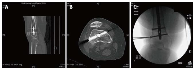
Figure 1 Preoperative imaging of femoral nail.
A 380 mm × 11 mm Synthes trochanteric femoral nail was in place from prior and now well-healed femoral neck fracture. Two 5 mm diameter distal locking screws were used. The distal-most locking screw was placed in the distal femur approximately 20 mm superior to the trochlear notch and oriented from posterolateral to anteromedial, in close proximity to the posterlateral femoral cortex and planned femoral tunnel. A: Sagittal; B: Axial CT images; C: Intraoperative fluoroscopic radiograph. CT: Computed tomography.
- Citation: Lacey M, Lamplot J, Walley KC, DeAngelis JP, Ramappa AJ. Technical note: Anterior cruciate ligament reconstruction in the presence of an intramedullary femoral nail using anteromedial drilling. World J Orthop 2017; 8(5): 379-384
- URL: https://www.wjgnet.com/2218-5836/full/v8/i5/379.htm
- DOI: https://dx.doi.org/10.5312/wjo.v8.i5.379


