Copyright
©The Author(s) 2017.
World J Orthop. May 18, 2017; 8(5): 372-378
Published online May 18, 2017. doi: 10.5312/wjo.v8.i5.372
Published online May 18, 2017. doi: 10.5312/wjo.v8.i5.372
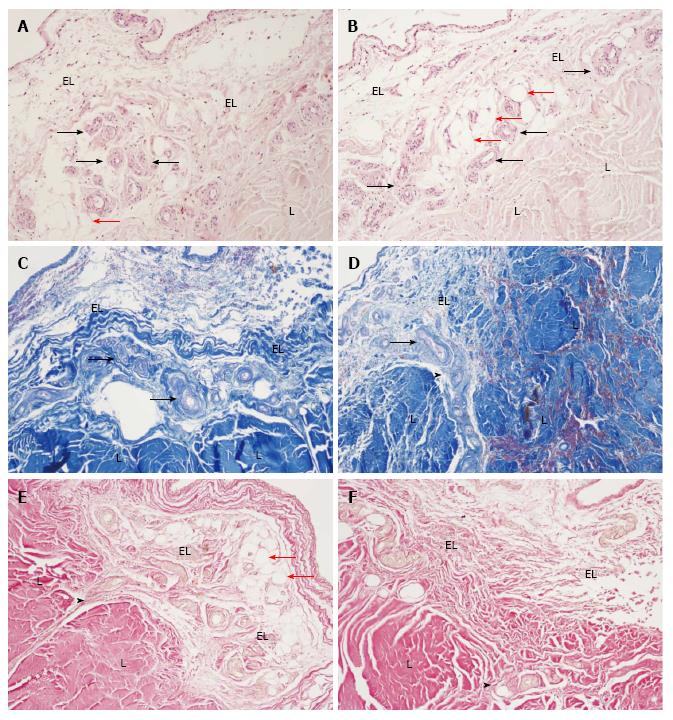
Figure 1 Normal morphology of the medial collateral ligament epiligament tissue in humans.
A and B: Normal morphology of the MCL EL tissue. Haematoxylin and eosin stain, × 200. EL: Epiligament; L: Ligament; red arrows: Adipocytes; arrows: Vessels in the EL tissue; C and D: Normal morphology of the MCL EL tissue. Mallory stain, × 200. EL: Epiligament; L: Ligament; arrows: Vessels in the EL tissue; arrow head: The EL extending into the endoligament; E and F: Normal morphology of the MCL EL tissue. Van Gieson’s stain, × 200. EL: Epiligament; L: Ligament; red arrows: Adipocytes; arrow head: The EL extending into the endoligament; MCL: Medial collateral ligament.
- Citation: Georgiev GP, Iliev A, Kotov G, Kinov P, Slavchev S, Landzhov B. Light and electron microscopic study of the medial collateral ligament epiligament tissue in human knees. World J Orthop 2017; 8(5): 372-378
- URL: https://www.wjgnet.com/2218-5836/full/v8/i5/372.htm
- DOI: https://dx.doi.org/10.5312/wjo.v8.i5.372


