Copyright
©The Author(s) 2017.
World J Orthop. Apr 18, 2017; 8(4): 301-309
Published online Apr 18, 2017. doi: 10.5312/wjo.v8.i4.301
Published online Apr 18, 2017. doi: 10.5312/wjo.v8.i4.301
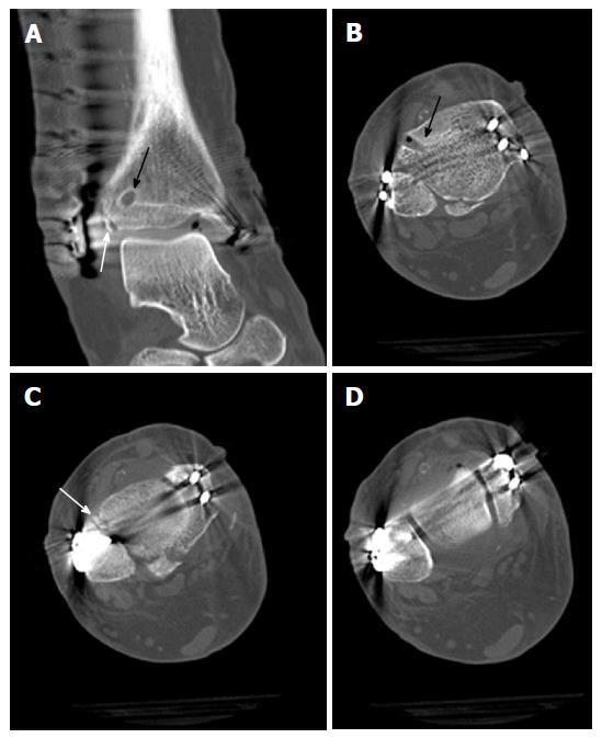
Figure 12 Postoperative computed tomography scans of the ankle from Figure 8 showing anatomic reduction of the tibial avulsion (white arrows) of the anterior tibiofibular ligament (A, C) as well as anatomic reduction of the ankle mortise (D); the tibial bone tunnel (black arrows) for the InternalBraceTM is clearly visible (A, B).
- Citation: Regauer M, Mackay G, Lange M, Kammerlander C, Böcker W. Syndesmotic InternalBraceTM for anatomic distal tibiofibular ligament augmentation. World J Orthop 2017; 8(4): 301-309
- URL: https://www.wjgnet.com/2218-5836/full/v8/i4/301.htm
- DOI: https://dx.doi.org/10.5312/wjo.v8.i4.301


