Copyright
©The Author(s) 2017.
World J Orthop. Apr 18, 2017; 8(4): 301-309
Published online Apr 18, 2017. doi: 10.5312/wjo.v8.i4.301
Published online Apr 18, 2017. doi: 10.5312/wjo.v8.i4.301
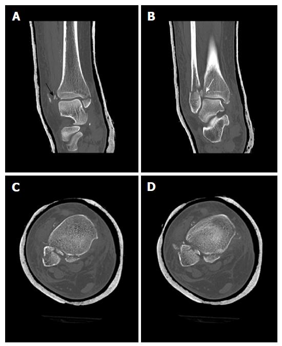
Figure 9 Computed tomography scans of the ankle from Figure 8 showing tibial avulsion of the anterior tibiofibular ligament with dislocation of a bone fragment (black arrow) too small for screw fixation (A), complete closed reduction was not possible due to a small bone fragment (white arrow) interposed between distal tibia and fibula (B), displaced avulsion of a small fragment of the posterolateral malleolus (C, D).
- Citation: Regauer M, Mackay G, Lange M, Kammerlander C, Böcker W. Syndesmotic InternalBraceTM for anatomic distal tibiofibular ligament augmentation. World J Orthop 2017; 8(4): 301-309
- URL: https://www.wjgnet.com/2218-5836/full/v8/i4/301.htm
- DOI: https://dx.doi.org/10.5312/wjo.v8.i4.301


