Copyright
©The Author(s) 2017.
World J Orthop. Apr 18, 2017; 8(4): 301-309
Published online Apr 18, 2017. doi: 10.5312/wjo.v8.i4.301
Published online Apr 18, 2017. doi: 10.5312/wjo.v8.i4.301
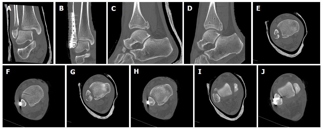
Figure 6 Syndesmotic InternalBraceTM for double stabilization.
Comparison of preoperative (A, C, E, G, I) and postoperative (B, D, F, H, J) CT scans. Note: anatomic positioning (F, H) and rotation (J) of the distal fibula and indirect anatomic reduction of the fracture of the posterior malleolus (D, F, H).
- Citation: Regauer M, Mackay G, Lange M, Kammerlander C, Böcker W. Syndesmotic InternalBraceTM for anatomic distal tibiofibular ligament augmentation. World J Orthop 2017; 8(4): 301-309
- URL: https://www.wjgnet.com/2218-5836/full/v8/i4/301.htm
- DOI: https://dx.doi.org/10.5312/wjo.v8.i4.301


