Copyright
©The Author(s) 2017.
World J Orthop. Mar 18, 2017; 8(3): 271-277
Published online Mar 18, 2017. doi: 10.5312/wjo.v8.i3.271
Published online Mar 18, 2017. doi: 10.5312/wjo.v8.i3.271
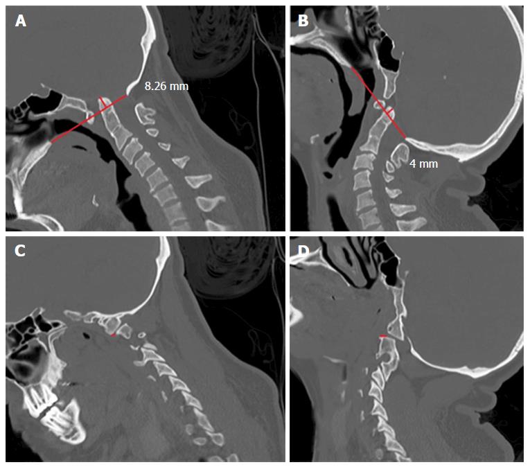
Figure 1 A 18-year-old woman with basilar invagination and tonsillar herniation of 7 mm.
She also had atlas assimilation. A: Sagittal computed tomography (CT) scan in flexion shows the tip of the odontoid 8.26 mm above the Chamberlain’s line; B: Sagittal CT scan in extended position shows the tip of the odontoid 4 mm above the Chamberlain’s line; C: Sagittal CT scan showing anterior dislocation of the facet joint of C1 over C2 facetary of 2 mm; D: Sagittal CT scan showing posterior dislocation of the C1 facet joint over the facet of C2 of 3 mm, ranging 5 mm in dynamic exam. This patient underwent a craniocervical fusion concomitant to the posterior fossa decompression.
- Citation: da Silva OT, Ghizoni E, Tedeschi H, Joaquim AF. Role of dynamic computed tomography scans in patients with congenital craniovertebral junction malformations. World J Orthop 2017; 8(3): 271-277
- URL: https://www.wjgnet.com/2218-5836/full/v8/i3/271.htm
- DOI: https://dx.doi.org/10.5312/wjo.v8.i3.271


