Copyright
©The Author(s) 2016.
World J Orthop. Jul 18, 2016; 7(7): 442-447
Published online Jul 18, 2016. doi: 10.5312/wjo.v7.i7.442
Published online Jul 18, 2016. doi: 10.5312/wjo.v7.i7.442
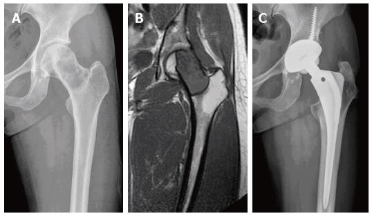
Figure 4 Radiographs of patient 1.
A: Radiograph of a 36-year-old woman shows an osteolytic lesion confined to femoral head and neck; B: Preoperative magnetic resonance imaging shows a low signal intensity lesion with well-defined surrounding rim; C: Postoperative radiograph at 6 years after total hip arthroplasty with extensive excision at the intertrochanteric level shows no evidence of local recurrence and implant loosening or osteolysis.
- Citation: Cho HS, Lee YK, Ha YC, Koo KH. Trochanter/calcar preserving reconstruction in tumors involving the femoral head and neck. World J Orthop 2016; 7(7): 442-447
- URL: https://www.wjgnet.com/2218-5836/full/v7/i7/442.htm
- DOI: https://dx.doi.org/10.5312/wjo.v7.i7.442


