Copyright
©The Author(s) 2016.
World J Orthop. Jul 18, 2016; 7(7): 406-417
Published online Jul 18, 2016. doi: 10.5312/wjo.v7.i7.406
Published online Jul 18, 2016. doi: 10.5312/wjo.v7.i7.406
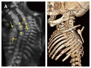
Figure 10 Rib fusions.
Grayscale 3D ultrasound image (A) of a 21 wk fetus demonstrate fusion of multiple left sided ribs (arrows) as well as multiple segmentation anomalies (arrowheads) resulting in congenital cervicothoracic kyphoscoliosis. Postnatal multidetector unenhanced computed tomography (B) of the thorax with volume rendering demonstrates bony fusion of multiple left sided ribs (arrows) as well as multiple segmentation anomalies and cervicothoracic dextroscoliosis.
- Citation: Upasani VV, Ketwaroo PD, Estroff JA, Warf BC, Emans JB, Glotzbecker MP. Prenatal diagnosis and assessment of congenital spinal anomalies: Review for prenatal counseling. World J Orthop 2016; 7(7): 406-417
- URL: https://www.wjgnet.com/2218-5836/full/v7/i7/406.htm
- DOI: https://dx.doi.org/10.5312/wjo.v7.i7.406


