Copyright
©The Author(s) 2016.
World J Orthop. Jul 18, 2016; 7(7): 406-417
Published online Jul 18, 2016. doi: 10.5312/wjo.v7.i7.406
Published online Jul 18, 2016. doi: 10.5312/wjo.v7.i7.406
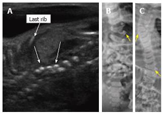
Figure 8 Thoracic hemivertebrae.
Parasagittal grayscale ultrasound image (A) of a 20 wk fetus acquired with a high frequency linear transducer demonstrate unpaired echogenic structures at the thoracolumbar junction representing hemivertebrae (arrows). Postnatal frontal radiographs of the same patient’s spine at 0 d (B) and 3 years of age (C) demonstrate left thoracolumbar and right midthoracic hemivertebrae.
- Citation: Upasani VV, Ketwaroo PD, Estroff JA, Warf BC, Emans JB, Glotzbecker MP. Prenatal diagnosis and assessment of congenital spinal anomalies: Review for prenatal counseling. World J Orthop 2016; 7(7): 406-417
- URL: https://www.wjgnet.com/2218-5836/full/v7/i7/406.htm
- DOI: https://dx.doi.org/10.5312/wjo.v7.i7.406


