Copyright
©The Author(s) 2016.
World J Orthop. Jul 18, 2016; 7(7): 406-417
Published online Jul 18, 2016. doi: 10.5312/wjo.v7.i7.406
Published online Jul 18, 2016. doi: 10.5312/wjo.v7.i7.406
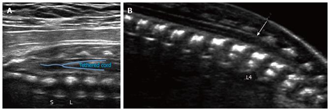
Figure 6 Spinal tether ultrasound.
Grayscale ultrasound image (A) of the lumbosacral spine in a 29 wk fetus demonstrates a low-lying conus medullaris, extending below the lumbosacral junction (L/S). No evidence of associated fatty filum terminale was seen in this case; B: Grayscale ultrasound image of the lumbosacral spine in a 21 wk fetus demonstrates a low-lying conus medullaris (arrow), extending below the inferior endplate of L3. No evidence of associated fatty filum terminale was seen in this case.
- Citation: Upasani VV, Ketwaroo PD, Estroff JA, Warf BC, Emans JB, Glotzbecker MP. Prenatal diagnosis and assessment of congenital spinal anomalies: Review for prenatal counseling. World J Orthop 2016; 7(7): 406-417
- URL: https://www.wjgnet.com/2218-5836/full/v7/i7/406.htm
- DOI: https://dx.doi.org/10.5312/wjo.v7.i7.406


