Copyright
©The Author(s) 2016.
World J Orthop. Dec 18, 2016; 7(12): 785-792
Published online Dec 18, 2016. doi: 10.5312/wjo.v7.i12.785
Published online Dec 18, 2016. doi: 10.5312/wjo.v7.i12.785
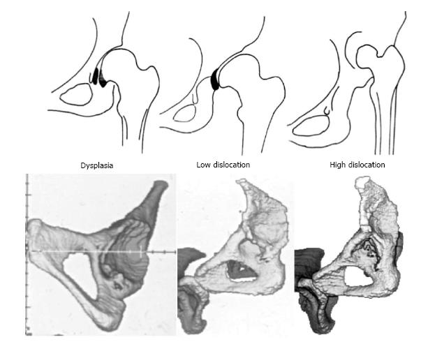
Figure 2 Drawings and three dimensional-computed tomography images of the 3 types of congenital hip disease in adults.
The black-colored areas in dysplasia represents the large osteophyte that covers the acetabular fossa and the medial marginal osteophyte of the femoral head (capital drop), and in low dislocation it the inferior part of the false acetabulum that is an osteophyte which begins at the level of the superior rim of the true acetabulum.
- Citation: Hartofilakidis G, Lampropoulou-Adamidou K. Lessons learned from study of congenital hip disease in adults. World J Orthop 2016; 7(12): 785-792
- URL: https://www.wjgnet.com/2218-5836/full/v7/i12/785.htm
- DOI: https://dx.doi.org/10.5312/wjo.v7.i12.785


