Copyright
©The Author(s) 2016.
World J Orthop. Oct 18, 2016; 7(10): 670-677
Published online Oct 18, 2016. doi: 10.5312/wjo.v7.i10.670
Published online Oct 18, 2016. doi: 10.5312/wjo.v7.i10.670
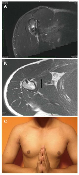
Figure 6 Magnetic resonance imaging at 6 mo follow up showing excellent continuity (arrow) and the bulk of pectoralis major muscle after repair (A, B); restoration of the anterior axillary fold after surgery (C).
- Citation: Joshi D, Jain JK, Chaudhary D, Singh U, Jain V, Lal A. Outcome of repair of chronic tear of the pectoralis major using corkscrew suture anchors by box suture sliding technique. World J Orthop 2016; 7(10): 670-677
- URL: https://www.wjgnet.com/2218-5836/full/v7/i10/670.htm
- DOI: https://dx.doi.org/10.5312/wjo.v7.i10.670


