Copyright
©The Author(s) 2016.
World J Orthop. Oct 18, 2016; 7(10): 638-649
Published online Oct 18, 2016. doi: 10.5312/wjo.v7.i10.638
Published online Oct 18, 2016. doi: 10.5312/wjo.v7.i10.638
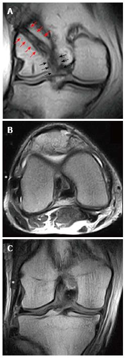
Figure 17 Magnetic resonance imaging evaluation of an anterior cruciate ligament reconstruction with a single-bundle plus lateral extra-articular plasty/augmentation using Gracilis and Semitendinosus Autograft.
In the coronal view, it is possible to identify the intra-articular part of the graft (black arrows) that continue proximally above the lateral femoral condyle (red arrows) in the “over-the-top” position (A). The lateral extra-articular plasty (asterisk) could be identified both in axial (B) and coronal view (C) beneath the iliotibial band, extending from the lateral femoral condyle to Gerdy’s tubercle.
- Citation: Grassi A, Bailey JR, Signorelli C, Carbone G, Wakam AT, Lucidi GA, Zaffagnini S. Magnetic resonance imaging after anterior cruciate ligament reconstruction: A practical guide. World J Orthop 2016; 7(10): 638-649
- URL: https://www.wjgnet.com/2218-5836/full/v7/i10/638.htm
- DOI: https://dx.doi.org/10.5312/wjo.v7.i10.638


