Copyright
©The Author(s) 2016.
World J Orthop. Oct 18, 2016; 7(10): 638-649
Published online Oct 18, 2016. doi: 10.5312/wjo.v7.i10.638
Published online Oct 18, 2016. doi: 10.5312/wjo.v7.i10.638
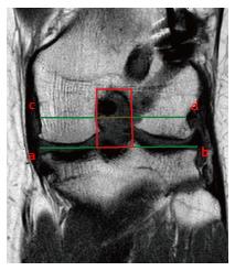
Figure 14 The femoral notch cross sectional area is measured as follows.
The coronal slice passing at the middle point of the Blumensaat Line is chosen. The width of the notch (red dotted line) is measured on a line passing through the popliteal groove (line c and d) parallel to the femoral joint surface (line a and b). The height of the notch is the distance between the joint surface and the top of the intercondylar notch. The cross sectional area (red box) is obtained multiplying the width (mm) by the height (mm).
- Citation: Grassi A, Bailey JR, Signorelli C, Carbone G, Wakam AT, Lucidi GA, Zaffagnini S. Magnetic resonance imaging after anterior cruciate ligament reconstruction: A practical guide. World J Orthop 2016; 7(10): 638-649
- URL: https://www.wjgnet.com/2218-5836/full/v7/i10/638.htm
- DOI: https://dx.doi.org/10.5312/wjo.v7.i10.638


