Copyright
©The Author(s) 2016.
World J Orthop. Oct 18, 2016; 7(10): 638-649
Published online Oct 18, 2016. doi: 10.5312/wjo.v7.i10.638
Published online Oct 18, 2016. doi: 10.5312/wjo.v7.i10.638
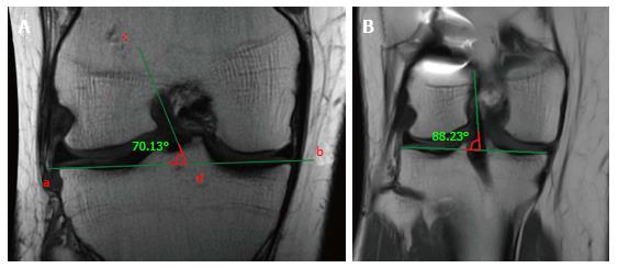
Figure 6 Measurement of the coronal obliquity of the graft.
The inclination is calculated measuring the angle between the tangent line to the tibial plateau (a and b) and the line which best defines the course of the intra-articular part of the graft (c and d). A high angle represents a vertical graft in the coronal plane (A). Vertically positioned graft, with an angle of 88°, far higher than the normal value < 75° (B).
- Citation: Grassi A, Bailey JR, Signorelli C, Carbone G, Wakam AT, Lucidi GA, Zaffagnini S. Magnetic resonance imaging after anterior cruciate ligament reconstruction: A practical guide. World J Orthop 2016; 7(10): 638-649
- URL: https://www.wjgnet.com/2218-5836/full/v7/i10/638.htm
- DOI: https://dx.doi.org/10.5312/wjo.v7.i10.638


