Copyright
©The Author(s) 2016.
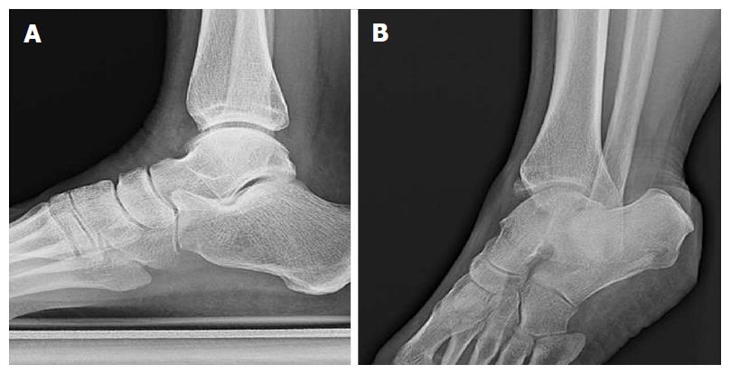
Figure 9 Standard lateral views about anterior tibial osteophytes.
Lateral plain radiograph of a right ankle with clinical suspicion of anteromedial impingement (A). An anteromedial radiographic view with 30 degrees of external rotation of the same ankle demonstrated an osteophyte on the anteromedial aspect of the distal tibia (B) that could not be appreciated on the lateral view.
- Citation: Walls RJ, Ross KA, Fraser EJ, Hodgkins CW, Smyth NA, Egan CJ, Calder J, Kennedy JG. Football injuries of the ankle: A review of injury mechanisms, diagnosis and management. World J Orthop 2016; 7(1): 8-19
- URL: https://www.wjgnet.com/2218-5836/full/v7/i1/8.htm
- DOI: https://dx.doi.org/10.5312/wjo.v7.i1.8


