Copyright
©The Author(s) 2016.
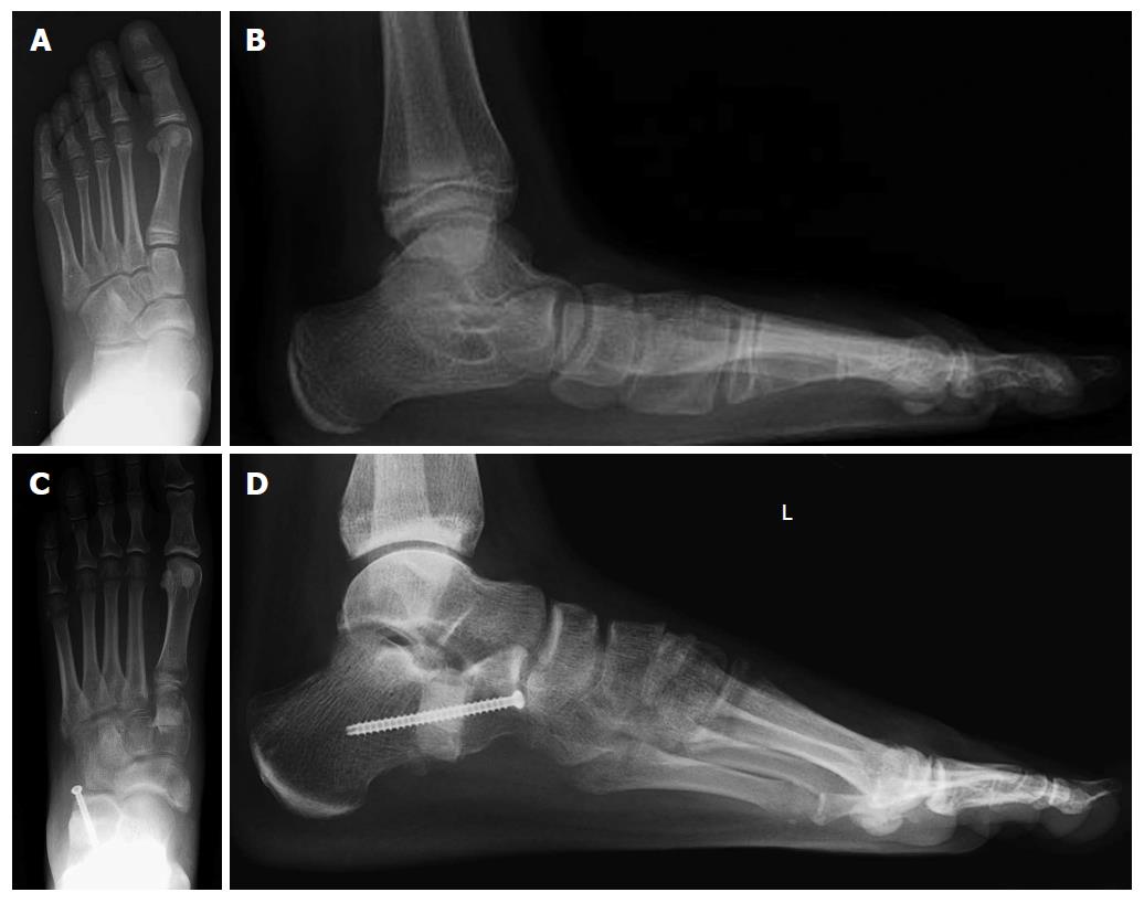
Figure 4 Preoperative anteroposterior (A) and lateral (B) views weightbearing X-rays in a child with a flexible flatfoot; on the anteroposterior view, note about 50% of talonavicular uncoverage; postoperative anteroposterior (C) and lateral (D) weightbearing views following a lateral column lengthening and cotton osteotomy.
Note the excellent correction of the talonavicular uncoverage (C) and Meary’s angle (D).
- Citation: Vulcano E, Maccario C, Myerson MS. How to approach the pediatric flatfoot. World J Orthop 2016; 7(1): 1-7
- URL: https://www.wjgnet.com/2218-5836/full/v7/i1/1.htm
- DOI: https://dx.doi.org/10.5312/wjo.v7.i1.1


