Copyright
©The Author(s) 2015.
World J Orthop. Aug 18, 2015; 6(7): 505-512
Published online Aug 18, 2015. doi: 10.5312/wjo.v6.i7.505
Published online Aug 18, 2015. doi: 10.5312/wjo.v6.i7.505
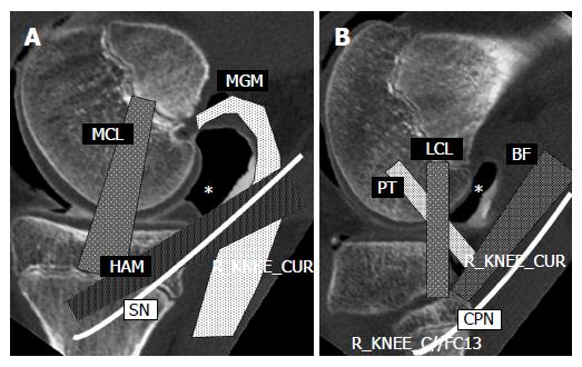
Figure 2 Sagittal images of double contrast arthrography at the levels of the medial (A) and lateral (B) femoral condyle of the knee.
Posteromedial (A) and posterolateral (B) sites are indicated by asterisks (*). The portal site is surrounded by important structures. MCL: Medial collateral ligament; MGM: Medial head of the gastrocnemius muscle; HAM: Hamstrings; SN: Saphenous nerve; LCL: Lateral collateral ligament; PT: Popliteus tendon; BF: Biceps femoris; CPN: Common peroneal nerve.
- Citation: Ohishi T, Takahashi M, Suzuki D, Matsuyama Y. Arthroscopic approach to the posterior compartment of the knee using a posterior transseptal portal. World J Orthop 2015; 6(7): 505-512
- URL: https://www.wjgnet.com/2218-5836/full/v6/i7/505.htm
- DOI: https://dx.doi.org/10.5312/wjo.v6.i7.505


