Copyright
©The Author(s) 2015.
World J Orthop. Mar 18, 2015; 6(2): 221-235
Published online Mar 18, 2015. doi: 10.5312/wjo.v6.i2.221
Published online Mar 18, 2015. doi: 10.5312/wjo.v6.i2.221
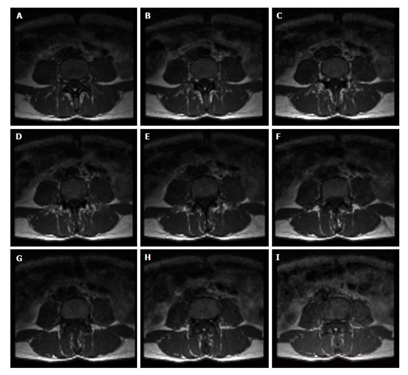
Figure 6 Set of images from magnetic resonance imaging spin-echo.
Nine axial images of the fourth lumbar vertebra obtained by magnetic resonance imaging multislice technique. Acquisition of the set of axial images of vertebral body to visualize the inner trabecular bone portion is performed from bottom (A: lower base) to top (I: upper base).
- Citation: Zaia A. Fractal lacunarity of trabecular bone and magnetic resonance imaging: New perspectives for osteoporotic fracture risk assessment. World J Orthop 2015; 6(2): 221-235
- URL: https://www.wjgnet.com/2218-5836/full/v6/i2/221.htm
- DOI: https://dx.doi.org/10.5312/wjo.v6.i2.221


