Copyright
©The Author(s) 2015.
World J Orthop. Mar 18, 2015; 6(2): 202-210
Published online Mar 18, 2015. doi: 10.5312/wjo.v6.i2.202
Published online Mar 18, 2015. doi: 10.5312/wjo.v6.i2.202
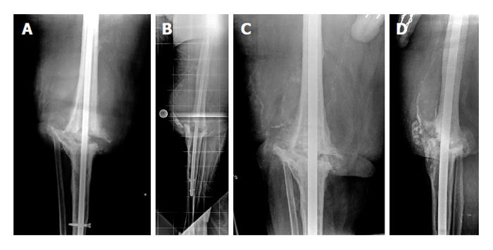
Figure 2 Anteroposterior and lateral view radiographs of the knee.
Anteroposterior (A) and lateral (B) view radiographs of the knee show a nonunion of a knee arthrodesis following insertion of an antegrade knee arthrodesis rod with inadequate tibial length. In this case, the intramedullary (IM) nail is shorter than what is typically used in such cases. The distal end of the nail should be 2 cm above the ankle joint. Also, this IM nail diameter is too small to provide a good canal fit. The IM diameter is measured, and exchange nailing is planned. Anteroposterior (C) and lateral (D) view radiographs of the knee after exchange nailing and bone grafting of the nonunion site. Exchange nailing to a larger diameter rod improves canal fit, thus providing additional stability to the nonunion site. The IM nail length was also increased so that the distal end of the nail was 2 cm above the ankle joint. Increasing both the length and the diameter improves the biomechanical stability of the nail and decreases motion at the nonunion site, thus promoting bony healing (reprinted with permission from the Rubin Institute for Advanced Orthopedics, Sinai Hospital of Baltimore).
- Citation: Wood JH, Conway JD. Advanced concepts in knee arthrodesis. World J Orthop 2015; 6(2): 202-210
- URL: https://www.wjgnet.com/2218-5836/full/v6/i2/202.htm
- DOI: https://dx.doi.org/10.5312/wjo.v6.i2.202


