Copyright
©The Author(s) 2015.
World J Orthop. Mar 18, 2015; 6(2): 202-210
Published online Mar 18, 2015. doi: 10.5312/wjo.v6.i2.202
Published online Mar 18, 2015. doi: 10.5312/wjo.v6.i2.202
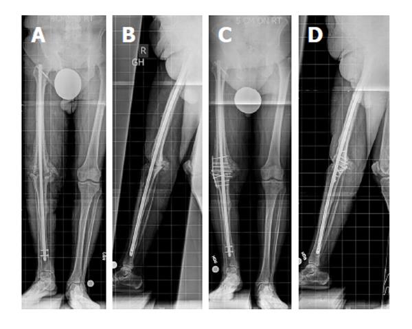
Figure 1 Radiographs of the knee.
Anteroposterior (A) and lateral (B) view radiographs of the knee show nonunion of a knee arthrodesis with a long intramedullary nail, without evidence of implant failure or loosening. The intramedullary (IM) nail diameter has a good diaphyseal canal fit but poor canal fit at the arthrodesis site and surrounding metaphysis, which decreases bending and rotational stability provided by the IM nail in this region. Decreased stability increases motion at the arthrodesis site, which can impede bony healing. Anteroposterior (C) and lateral (D) view radiographs of the knee after supplemental plate fixation around the existing knee fusion nail and bone grafting of the nonunion site. Supplemental plate fixation provides additional stability at the nonunion site, which promotes bony healing and union of the knee arthrodesis. Bicortical screw fixation is achieved by aiming slightly posterior to the IM nail. Note the motion at the distal interlocking screws is now visible; however, the knee arthrodesis site has progressive consolidation (reprinted with permission from the Rubin Institute for Advanced Orthopedics, Sinai Hospital of Baltimore).
- Citation: Wood JH, Conway JD. Advanced concepts in knee arthrodesis. World J Orthop 2015; 6(2): 202-210
- URL: https://www.wjgnet.com/2218-5836/full/v6/i2/202.htm
- DOI: https://dx.doi.org/10.5312/wjo.v6.i2.202


