Copyright
©The Author(s) 2015.
World J Orthop. Nov 18, 2015; 6(10): 829-837
Published online Nov 18, 2015. doi: 10.5312/wjo.v6.i10.829
Published online Nov 18, 2015. doi: 10.5312/wjo.v6.i10.829
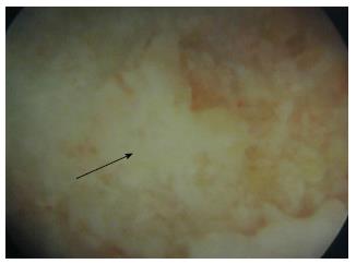
Figure 1 Endoscopic view of the osteonecrotic lesion.
View obtained from the endoscopy through the canal. The posterior aspect of the osteonecrotic lesion is seen at the center of the image (arrow) as a white non-vascularized area, surrounded by normal purple-coloured bone (vascularized bone).
- Citation: Pakos EE, Megas P, Paschos NK, Syggelos SA, Kouzelis A, Georgiadis G, Xenakis TA. Modified porous tantalum rod technique for the treatment of femoral head osteonecrosis. World J Orthop 2015; 6(10): 829-837
- URL: https://www.wjgnet.com/2218-5836/full/v6/i10/829.htm
- DOI: https://dx.doi.org/10.5312/wjo.v6.i10.829


