Copyright
©2014 Baishideng Publishing Group Inc.
World J Orthop. Nov 18, 2014; 5(5): 614-622
Published online Nov 18, 2014. doi: 10.5312/wjo.v5.i5.614
Published online Nov 18, 2014. doi: 10.5312/wjo.v5.i5.614
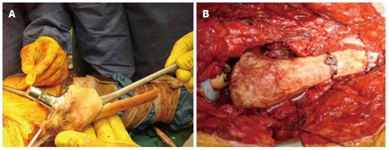
Figure 8 The allograft is reamed and broached and a long stem is cemented at the back table.
A: Intraoperative picture demonstrating the allograft-prosthesis composite preparation. An allograft of appropriate size is osteotomized at the desired subtrochanteric level in order to match the bony defect of the proximal femur. The allograft is reamed and broached and a long stem is cemented at the back table; B: Intraoperative picture showing the allograft-prosthesis composite with a lateral sleeve that offers a wide area of bone contact with the distal host femur. Circlage cables are used to secure the allograft-host bone fixation.
- Citation: Sakellariou VI, Babis GC. Management bone loss of the proximal femur in revision hip arthroplasty: Update on reconstructive options. World J Orthop 2014; 5(5): 614-622
- URL: https://www.wjgnet.com/2218-5836/full/v5/i5/614.htm
- DOI: https://dx.doi.org/10.5312/wjo.v5.i5.614


