Copyright
©2014 Baishideng Publishing Group Inc.
World J Orthop. Nov 18, 2014; 5(5): 614-622
Published online Nov 18, 2014. doi: 10.5312/wjo.v5.i5.614
Published online Nov 18, 2014. doi: 10.5312/wjo.v5.i5.614
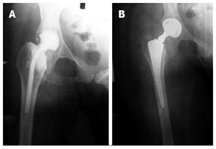
Figure 3 Anteroposterior radiograph of bone loss.
A: Anteroposterior (AP) radiograph of the right hip showing minimal bone loss of the proximal metaphyseal bone secondary to periprosthetic hip infection. A antibiotic cement spacer was implanted after irrigation and debridement during the first stage of a two-stage exchange arthroplasty; B: AP radiograph of the same hip after the second stage. The metaphyseal bone loss was minimal and a cementless primary stems with common length and geometry was used.
- Citation: Sakellariou VI, Babis GC. Management bone loss of the proximal femur in revision hip arthroplasty: Update on reconstructive options. World J Orthop 2014; 5(5): 614-622
- URL: https://www.wjgnet.com/2218-5836/full/v5/i5/614.htm
- DOI: https://dx.doi.org/10.5312/wjo.v5.i5.614


