Copyright
©2014 Baishideng Publishing Group Inc.
World J Orthop. Nov 18, 2014; 5(5): 614-622
Published online Nov 18, 2014. doi: 10.5312/wjo.v5.i5.614
Published online Nov 18, 2014. doi: 10.5312/wjo.v5.i5.614
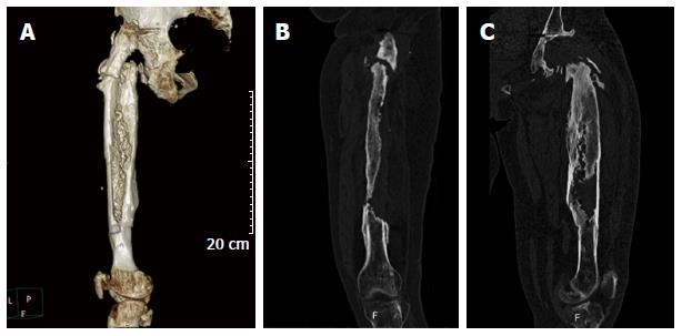
Figure 1 Computed tomography scan images with metallic artifact subtraction and three-dimensional reconstruction consist a useful tool for precise assessment of the amount of bone loss and the specific variations of the femoral anatomy preoperatively.
A: Computed tomography scan image of the left femur (posterior projection) with metallic artifact subtraction and three-dimensional reconstruction showing precisely the amount of bone loss of the proximal part of the femur; B: Coronal; C: Sagittal view of the same case.
- Citation: Sakellariou VI, Babis GC. Management bone loss of the proximal femur in revision hip arthroplasty: Update on reconstructive options. World J Orthop 2014; 5(5): 614-622
- URL: https://www.wjgnet.com/2218-5836/full/v5/i5/614.htm
- DOI: https://dx.doi.org/10.5312/wjo.v5.i5.614


