Copyright
©2014 Baishideng Publishing Group Inc.
World J Orthop. Nov 18, 2014; 5(5): 574-584
Published online Nov 18, 2014. doi: 10.5312/wjo.v5.i5.574
Published online Nov 18, 2014. doi: 10.5312/wjo.v5.i5.574
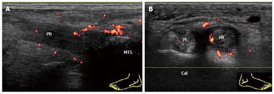
Figure 14 Involvement of peroneal brevis tendon in psoriatic arthritis.
A: Enthesitis of the PB in psoriatic arthritis. Longitudinal power Doppler sonogram of the PB shows intratendinous hypoechoic change and loss of the fibrillar echoes with hyperemia adjacent to the insertion into the base of MT5. Cortical irregularities of the bone at the insertion are also depicted; B: Tenosynovitis of the contralateral PB in the same patient. Transverse power Doppler sonogram at the level of the peroneal tubercle of the Cal shows thickening and hyperemia of the PB tendon sheath. PL: Peroneus longus; PB: Peroneus brevis; MT5: The fifth metatarsal bone; Cal: Calcaneus.
- Citation: Suzuki T. Power Doppler ultrasonographic assessment of the ankle in patients with inflammatory rheumatic diseases. World J Orthop 2014; 5(5): 574-584
- URL: https://www.wjgnet.com/2218-5836/full/v5/i5/574.htm
- DOI: https://dx.doi.org/10.5312/wjo.v5.i5.574


