Copyright
©2014 Baishideng Publishing Group Inc.
World J Orthop. Nov 18, 2014; 5(5): 574-584
Published online Nov 18, 2014. doi: 10.5312/wjo.v5.i5.574
Published online Nov 18, 2014. doi: 10.5312/wjo.v5.i5.574
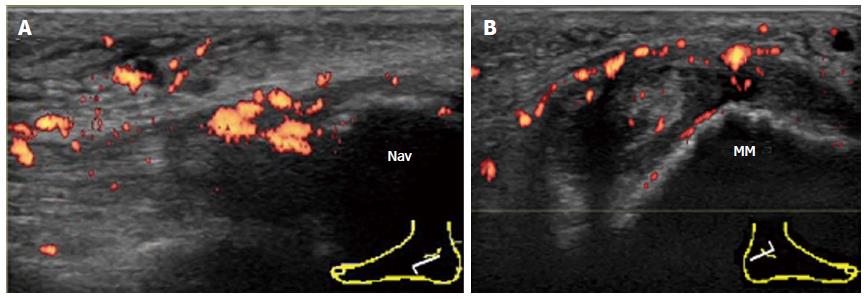
Figure 11 Involvement of the tibialis posterior tendon in psoriatic arthritis.
A: Enthesitis of the TP at the navicular insertion in psoriatic arthritis. Longitudinal power Doppler sonogram of TP shows intratendinous hypoechoic change and loss of the fibrillar echoes with hyperemia adjacent to the insertion into thenavicular bone. Cortical irregularities of the navicular bone at the insertion are also depicted; B: Tenosynovitis of the contralateral TP in the same patient. Transverse power Doppler sonogram at the level of the tip of the medial malleolus shows thickening and hyperemia of both the tendon sheath and the flexor retinaculum. Cortical irregularities of the medial malleolus are also depicted. Nav: Navicular bone; MM: Medial malleolus; TP: Tibialis posterior.
- Citation: Suzuki T. Power Doppler ultrasonographic assessment of the ankle in patients with inflammatory rheumatic diseases. World J Orthop 2014; 5(5): 574-584
- URL: https://www.wjgnet.com/2218-5836/full/v5/i5/574.htm
- DOI: https://dx.doi.org/10.5312/wjo.v5.i5.574


