Copyright
©2014 Baishideng Publishing Group Inc.
World J Orthop. Nov 18, 2014; 5(5): 574-584
Published online Nov 18, 2014. doi: 10.5312/wjo.v5.i5.574
Published online Nov 18, 2014. doi: 10.5312/wjo.v5.i5.574
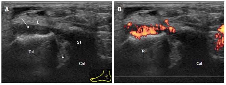
Figure 5 Synovitis of the posterior subtalar joint in rheumatoid arthritis.
Coronal grey-scale (A) and power Doppler (B) sonogram of the medial facet (arrowhead) of the PSTJ. PD-signal-positive proliferated synovium (arrow) of the PSTJ stretches cranially along the tibiocalcaneal ligament (L). Tal: Talus; Cal: Calcaneus; ST: Sustentaculum tali; PSTJ: Posterior subtalar joint.
- Citation: Suzuki T. Power Doppler ultrasonographic assessment of the ankle in patients with inflammatory rheumatic diseases. World J Orthop 2014; 5(5): 574-584
- URL: https://www.wjgnet.com/2218-5836/full/v5/i5/574.htm
- DOI: https://dx.doi.org/10.5312/wjo.v5.i5.574


