Copyright
©2014 Baishideng Publishing Group Inc.
World J Orthop. Jul 18, 2014; 5(3): 272-282
Published online Jul 18, 2014. doi: 10.5312/wjo.v5.i3.272
Published online Jul 18, 2014. doi: 10.5312/wjo.v5.i3.272
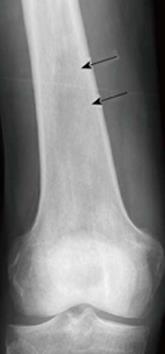
Figure 1 X-ray of an osseous myeloma lesion.
Conventional X-ray of the right femoral bone showing an osteolytic lesion (arrows) representing an osseous myeloma manifestation.
- Citation: Derlin T, Bannas P. Imaging of multiple myeloma: Current concepts. World J Orthop 2014; 5(3): 272-282
- URL: https://www.wjgnet.com/2218-5836/full/v5/i3/272.htm
- DOI: https://dx.doi.org/10.5312/wjo.v5.i3.272


