Copyright
©2014 Baishideng Publishing Group Inc.
World J Orthop. Jul 18, 2014; 5(3): 171-179
Published online Jul 18, 2014. doi: 10.5312/wjo.v5.i3.171
Published online Jul 18, 2014. doi: 10.5312/wjo.v5.i3.171
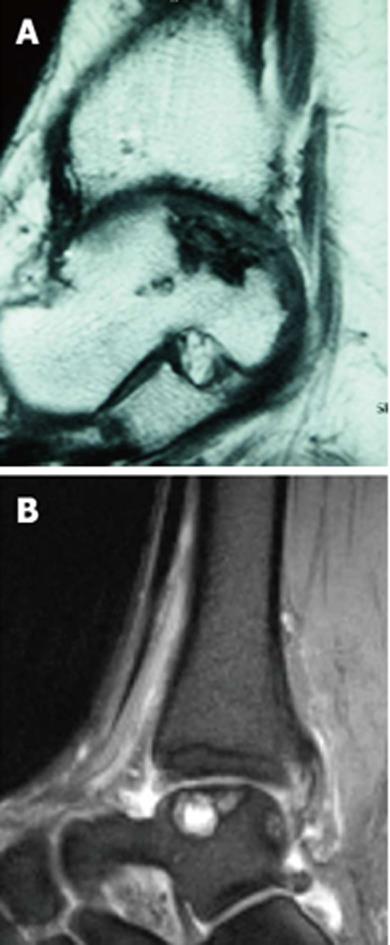
Figure 5 Sagittal T1 and T2-weighted magnetic resonance imaging scan.
A: Sagittal T1-weighted magnetic resonance imaging scan demonstrating deep osteochondral defect of the posterior aspect of the talus; B: Sagittal T2-weighted magnetic resonance imaging scan showing the several cystic lesions of the talus in addition to an osteochondral defect.
- Citation: Baums MH, Schultz W, Kostuj T, Klinger HM. Cartilage repair techniques of the talus: An update. World J Orthop 2014; 5(3): 171-179
- URL: https://www.wjgnet.com/2218-5836/full/v5/i3/171.htm
- DOI: https://dx.doi.org/10.5312/wjo.v5.i3.171


