Copyright
©2014 Baishideng Publishing Group Co.
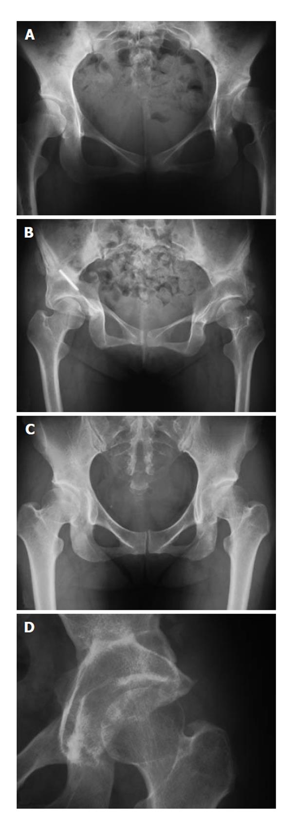
Figure 2 Anteroposterior X-ray images of cases 1, 2 and 5.
A: Pre-operative anteroposterior (AP) X-ray images of cases 1 and 2 showing pre-osteoarthritis in a patient with bilateral developmental dysplasia of the hip (DDH); B: Post-operative AP X-ray image at the final follow-up examination. Three 3.0 mm K-wires were used for fixation of the acetabulum. One of the K-wire was remained at the right hip; C: Pre-operative AP X-ray image showing pre-osteoarthritis in a patient with bilateral DDH; D: Post-operative AP X-ray image at the final follow-up examination.
- Citation: Mimura T, Mori K, Kawasaki T, Imai S, Matsusue Y. Triple pelvic osteotomy: Report of our mid-term results and review of literature. World J Orthop 2014; 5(1): 14-22
- URL: https://www.wjgnet.com/2218-5836/full/v5/i1/14.htm
- DOI: https://dx.doi.org/10.5312/wjo.v5.i1.14


