Copyright
©2013 Baishideng Publishing Group Co.
World J Orthop. Oct 18, 2013; 4(4): 327-332
Published online Oct 18, 2013. doi: 10.5312/wjo.v4.i4.327
Published online Oct 18, 2013. doi: 10.5312/wjo.v4.i4.327
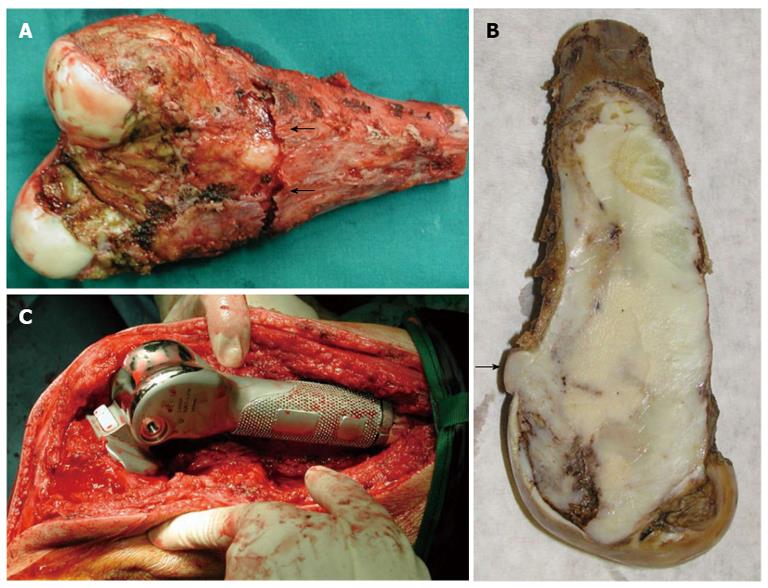
Figure 4 Open biopsy was performed obtaining bone specimens from the outer lateral condyle with 3 cm × 2 cm × 1 cm total dimensions.
A: Excised specimen of the distal 15.5 cm of the femur. Compressive fracture of the posterior cortex (arrows); B: The tumor arose within the medullary cavity and extended from the cartilage surface to 3 cm distance from the excision line. The mass clearly breaches the cortex (arrow); C: Hinged custom made total knee arthroplasty (Howmedica Modular Resection System, Stryker Howmedica Osteonics, Inc).
- Citation: Vasiliadis HS, Arnaoutoglou C, Plakoutsis S, Doukas M, Batistatou A, Xenakis TA. Low-grade central osteosarcoma of distal femur, resembling fibrous dysplasia. World J Orthop 2013; 4(4): 327-332
- URL: https://www.wjgnet.com/2218-5836/full/v4/i4/327.htm
- DOI: https://dx.doi.org/10.5312/wjo.v4.i4.327


