Copyright
©2013 Baishideng Publishing Group Co.
World J Orthop. Oct 18, 2013; 4(4): 327-332
Published online Oct 18, 2013. doi: 10.5312/wjo.v4.i4.327
Published online Oct 18, 2013. doi: 10.5312/wjo.v4.i4.327
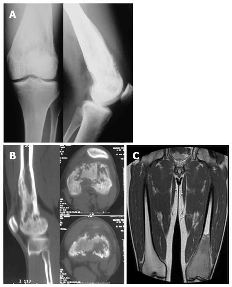
Figure 1 Imaging of the lesion at the patient admission.
A: Lytic lesion of the distal femur is seen on radiographs; B: Computed tomography scan of distal femur showing the erosion of the bone with focal cortical destruction; C: T1-weighted magnetic resonance imaging demonstrating the extent of the lesion with areas of cortical destruction and periostic reaction.
- Citation: Vasiliadis HS, Arnaoutoglou C, Plakoutsis S, Doukas M, Batistatou A, Xenakis TA. Low-grade central osteosarcoma of distal femur, resembling fibrous dysplasia. World J Orthop 2013; 4(4): 327-332
- URL: https://www.wjgnet.com/2218-5836/full/v4/i4/327.htm
- DOI: https://dx.doi.org/10.5312/wjo.v4.i4.327


