Copyright
©2011 Baishideng Publishing Group Co.
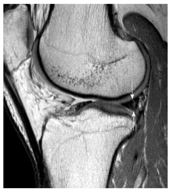
Figure 16 Peripheral vertical tear in a patient with complete anterior cruciate ligament tear.
Sagittal intermediate-weighted magnetic resonance image demonstrates a peripheral vertical tear (white arrows) extending of the posterior horn of lateral meniscus. This kind of peripheral vertical tear can be easily missed and is frequently associated with anterior cruciate ligament tear.
- Citation: Ng WHA, Griffith JF, Hung EHY, Paunipagar B, Law BKY, Yung PSH. Imaging of the anterior cruciate ligament. World J Orthop 2011; 2(8): 75-84
- URL: https://www.wjgnet.com/2218-5836/full/v2/i8/75.htm
- DOI: https://dx.doi.org/10.5312/wjo.v2.i8.75


