Copyright
©2011 Baishideng Publishing Group Co.
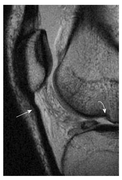
Figure 11 Patellar buckling sign and lateral femoral notch sign.
A 37-year-old man who suffered knee injury. Osteochondral injury and patellar buckling sign. Sagittal intermediate-weighted magnetic resonance image demonstrates a deep depression of the middle portion of the lateral femoral condyle (curved white arrow). Normal condylopatellar sulcus should be smaller than 1.5 mm. Notch depth between 1 and 2 mm is suggestive and over 2 mm is diagnostic of anterior cruciate ligament tear. Buckling of proximal patellar tendon (white arrow) also indicates the underlying anterior cruciate ligament tear.
- Citation: Ng WHA, Griffith JF, Hung EHY, Paunipagar B, Law BKY, Yung PSH. Imaging of the anterior cruciate ligament. World J Orthop 2011; 2(8): 75-84
- URL: https://www.wjgnet.com/2218-5836/full/v2/i8/75.htm
- DOI: https://dx.doi.org/10.5312/wjo.v2.i8.75


